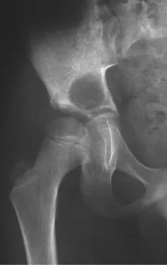
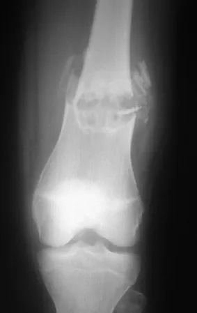
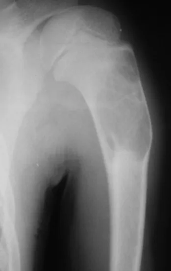
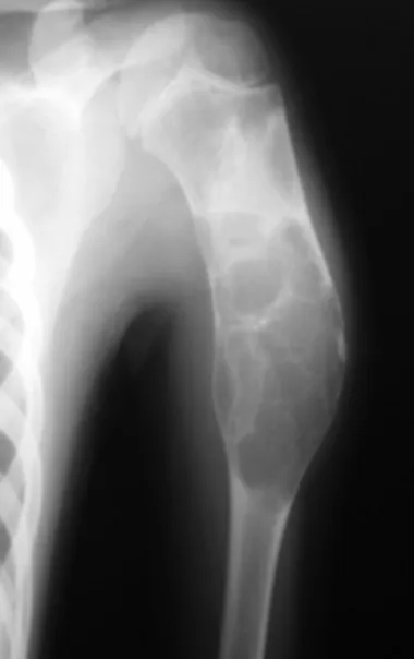
This digital library houses the book on Oncology and Orthopedic Oncosurgery.

It includes academic lectures, presentations from national and international congresses, published papers, case discussions, performed surgical procedures, and proprietary techniques developed.

The digital format was chosen because the web allows the inclusion of texts with numerous visual resources, such as images and videos, which would not be possible in a printed book.

The content is intended for students, healthcare professionals, and the general public interested in the field.

Pathological Fractures in Children

Fraturas em Crianças
[auto_translate_button]

Fractures in Children with Preexisting Bone Conditions

When we talk about “pathological fractures in children”, it is essential to clarify that the fracture itself is not pathological, but rather the bone can present a series of changes, such as structural, metabolic, dysplastic or infectious. Therefore, it is more accurate to refer to the bone as pathological, not the fracture itself. Within this context, our focus will be on fractures that occur in children with pre-existing bone conditions.

This chapter aims to address more specifically the fractures that develop in children with such conditions. It is important to highlight that the scope of this topic is vast, which leads us to define the topics to be discussed.

We decided not to include fractures related to infectious processes or metabolic disorders, such as rickets or osteopsatirosis, in this chapter. Instead, our focus will be on stress fractures, considering the differential diagnosis, as well as those arising from pre-existing tumoral or pseudo-tumorous bone lesions.

Stress fractures are particularly relevant due to their nature and challenges associated with diagnosis and treatment. Furthermore, fractures resulting from tumor or pseudo-tumor bone lesions require a specialized approach to ensure appropriate management and the best possible prognosis.

Therefore, in outlining this chapter, we seek to provide a comprehensive view of fractures in children with pre-existing bone conditions, highlighting the most relevant aspects for their understanding and clinical management.

  • Benign Bone Tumors:

Among the benign tumor lesions of childhood, which can most frequently cause fractures, we highlight osteoblastoma and chondroblastoma.

Osteoblastoma – 

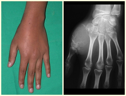
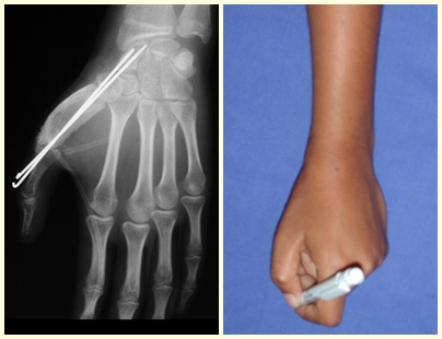
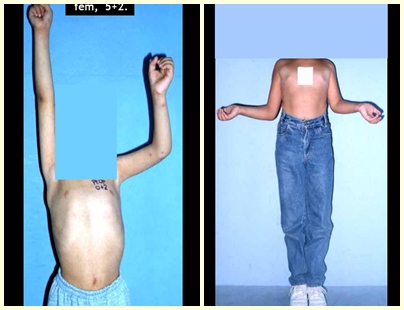
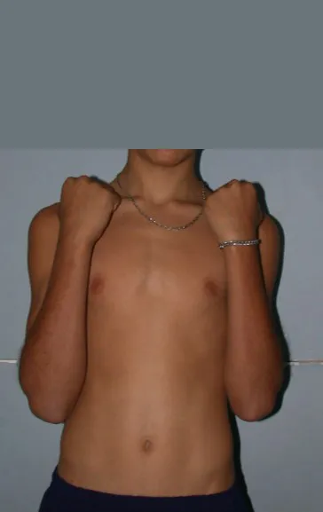
Osteoblastoma is a locally aggressive bone tumor that in long bones has a metaphyseal location, initially cortical and eccentric. This injury, as it is locally aggressive, with great destruction of the bone framework, causes micro fractures, due to erosion of the bone cortex (figs. 1 and 2). The progressive destruction of the cortex predisposes to complete fracture, when the involvement exceeds fifty percent of the bone circumference. The fracture of this lesion facilitates local dissemination, making oncological treatment difficult, which requires elaborate reconstructions and there is a limitation in functional recovery (Figs. 3 and 4). 

Figura 1 – aspecto clínico de osteoblastoma do primeiro metacarpeano. Fig. 2 – lesão insuflativa, com erosão da cortical e microfraturas.
Figure 1 – clinical appearance of first metacarpal osteoblastoma. Fig. 2 – Insufflation injury, with cortical erosion and microfractures.
Figura 3 – agressividade local com grande destruição da estrutura óssea, sendo necessária a reconstrução com enxerto autólogo do ilíaco, apesar da baixa idade. Fig. 4 – aspecto clínico e funcional, pós-operatório.
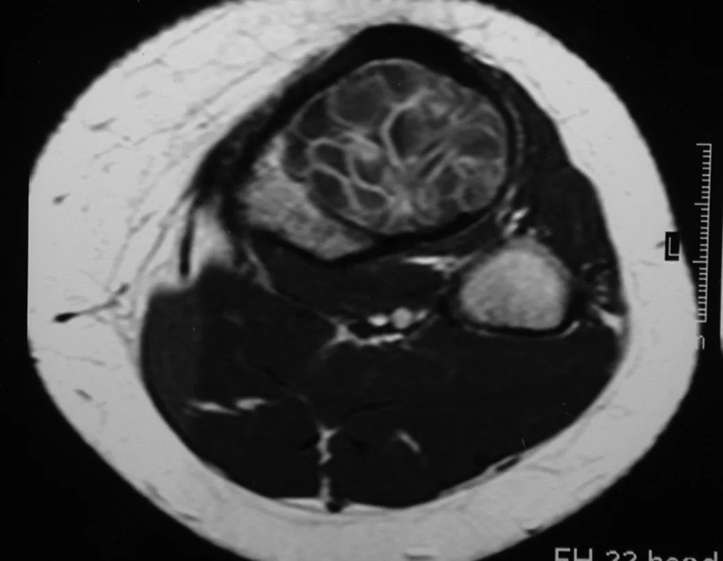
Figure 3 – local aggressiveness with great destruction of the bone structure, requiring reconstruction with an autologous iliac graft, despite young age. Fig. 4 – clinical and functional appearance, post-operatively.
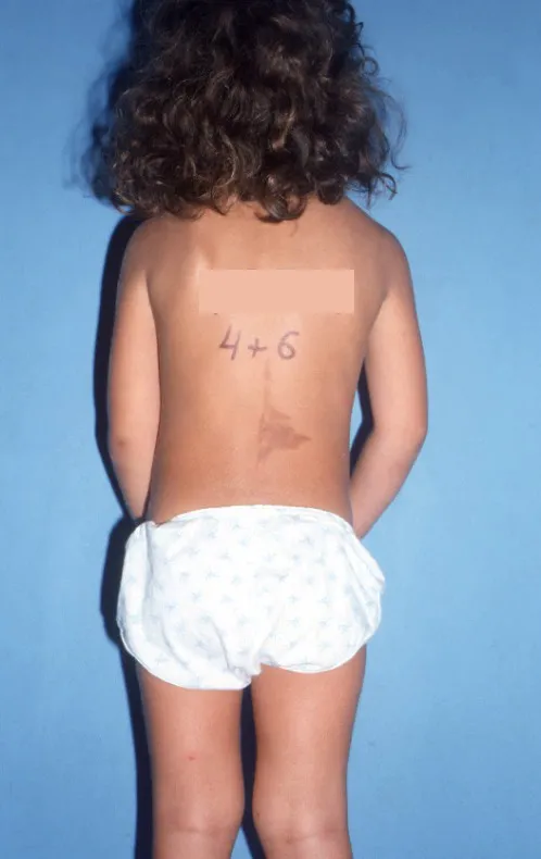
Osteoblastoma of the spinal pedicle can cause analgesic scoliosis, due to the pain of the tumor process or the fracture (fig. 5 and 6).
Figura 5 – escoliose antálgica devido à osteoblastoma. Fig. 6 – fratura do pedículo pelo tumor ósseo.
Figure 5 – Antalgic scoliosis due to osteoblastoma. Fig. 6 – fracture of the pedicle caused by the bone tumor.

Chondroblastoma –

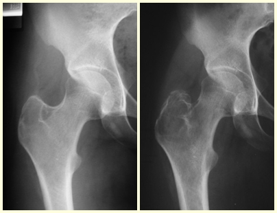
Chondroblastoma affects the epiphyseal region of growing long bones (figs. 7 and 8) and, less frequently, the apophyseal portion (figs. 9 and 10).

Figura 1 – condroblastoma epifisário do fêmur.
Figure 1 – Epiphyseal chondroblastoma of the femur.
Figura 2– erosão da cartilagem articular.
Figure 2– erosion of the articular cartilage.
3 – condroblastoma da apófise do grande trocânter. Fig. 4 – aumento significativo da lesão, com fratura arrancamento do grande trocânter.
3 – chondroblastoma of the apophysis of the greater trochanter. Fig. 4 – significant increase in the lesion, with a tearing fracture of the greater trochanter.

This bone tumor causes resorption of the epiphysis (or apophysis), erosion of the bone cortex and joint invasion, leading to arthralgia, which can cause deformity and joint subsidence fracture.

The treatment of both osteoblastoma and chondroblastoma is surgical and must be carried out as soon as possible, as these lesions, despite being histologically benign, quickly progress to destruction of the local bone framework.

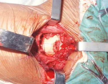
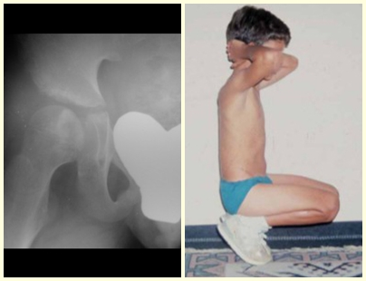
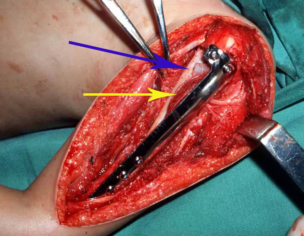
The best indication to avoid local recurrence is segmental resection. However, due to the articular location of the chondroblastoma, it is preferable to provide adequate surgical access to each region, as in this example that affects the posteromedial region of the femoral head (fig. 11), to perform careful intra-lesional curettage, followed by local adjuvant , such as phenol, liquid nitrogen or electrothermia (fig. 12), to subsequently fill the cavity with an autologous bone graft, restoring the anatomy of the region (fig. 13) and reestablishing function (figs 14 and 15). 

Figura 7 – via de acesso à região póstero-medial da cabeça femoral.
Figure 7 – access route to the posteromedial region of the femoral head.
Figura 8 – curetagem intralesional, seguida de eletrotermia.
Figure 8 – Intralesional curettage, followed by electrothermia.
Figura 9 – enxerto ósseo autólogo já integrado. Fig. 10 – Função de flexão com carga dos quadris.
Figure 9 – autologous bone graft already integrated. Fig. 10 – Load-bearing flexion function of the hips.
Figura 11 Abdução do quadril, após um ano da cirurgia.
Figure 11 Hip abduction, one year after surgery.
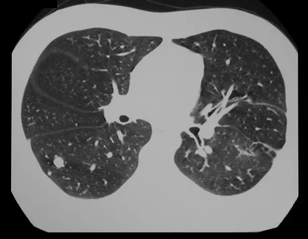
Chondroblastoma, despite being a benign lesion, in addition to local recurrence, can evolve into pulmonary metastases (fig. 16 and 17) which remain histologically benign, making the indication of additional chemotherapy controversial.
Figura 12 – radiografia do tórax, com múltiplos nódulos.
Figure 12 – chest x-ray, with multiple nodules.
Figura 13 – tomografia do tórax, com treze anos de evolução.
Figure 13 – Chest tomography, with thirteen years of evolution.

 In our experience we had two cases of osteoblastoma and one case of chondroblastoma with secondary lung disease. In this case of chondroblastoma, thoracotomy was performed and numerous pulmonary nodules were found, which persist to this day. This patient, at the time of diagnosis of metastases, presented with hypertrophic pulmonary osteopathy. He did not undergo any complementary treatment and is asymptomatic to this day, thirteen years later (fig. 18 and ’19) and fifteen years after surgery (fig. 20 and 21).

Figura 14 – Abdução apos 8 anos. Fig. 15 – Fexão com carga após 13 anos.
Figure 14 – Abduction after 8 years. Fig. 15 – Loaded bending after 13 years.
Figura 16 – Abdução após 15 anos. Fig. 17 Flexão com carga após quinze anos da cirurgia.
Figure 16 – Abduction after 15 years. Fig. 17 Flexion with weight fifteen years after surgery.
  • Malignant Bone Tumors:

The most common malignant bone neoplasms in childhood are osteosarcoma and Ewing’s sarcoma, which must be diagnosed at the onset of symptoms, as they cause pain and a palpable tumor and need to be treated early.

Osteosarcoma –

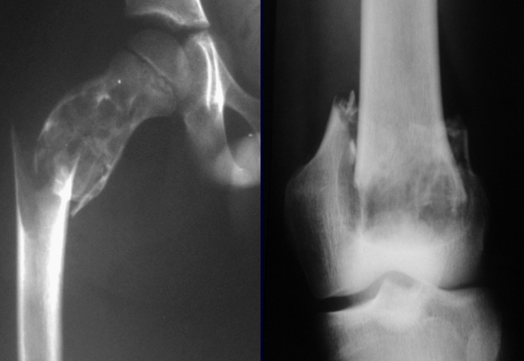
In our country, it is not uncommon for osteosarcoma to present with a fracture at diagnosis (figs. 22 and 23).  

Figura 22 – osteossarcoma da metáfise distal do fêmur. Fig. 23 – fratura e disseminação local ao diagnóstico.
Figure 22 – Osteosarcoma of the distal metaphysis of the femur. Fig. 23 – fracture and local spread at diagnosis.
In these situations, local oncological control may require ablative surgery, with Van-Ness gyroplasty (figs. 24 and 25) being an alternative to be considered.
Figura 24 – incisão rombóide para a cirurgia de Van-Ness.
Figure 24 – rhomboid incision for Van-Ness surgery.
Figura 25 – ressecção ampla, com margem oncológica, preservando-se o feixe vasculo-nervoso.
Figure 25 – wide resection, with oncological margin, preserving the vascular-nervous bundle.
This surgery is an interim amputation that changes the function of the ankle. This undergoes a 180 degree rotation and will act as if it were the knee, with the aim of transforming an amputation at thigh level into a “below the knee” amputation. Functionally, it acts as if it were an amputation of the leg, with the terminal support of the calcaneus and without the need for a mechanical “knee” (figs. 26 and 27). Special orthoses need to be made to fit the patient (fig. 28 and 29). There is a need for social and psychological support for the success of this procedure, which is little accepted and therefore rarely recommended in our country.
Figura 26 – giroplastia de Van-Ness, apoio terminal no calcâneo. Fig. 27 – a contratura dos ísquio tibiais, suturados nos flexores dorsais do tornozelo, realizam a flexão.
Figure 26 – Van-Ness gyroplasty, terminal support on the calcaneus. Fig. 27 – the contracture of the tibial ischium, sutured to the ankle's dorsal flexors, performs flexion.
Figura 28 – a contratura do quadríceps, suturado no tendão calcâneo, realiza a extensão do “neo joelho”. Fig. 29 – órteses especiais para adaptação. Há necessidade de fisioterapia especializada e apoio psicológico e social ao paciente e aos familiares.
Figure 28 – quadriceps contracture, sutured to the Achilles tendon, extends the “neo knee”. Fig. 29 – special orthoses for adaptation. There is a need for specialized physiotherapy and psychological and social support for the patient and family.

Ewing sarcoma – 

Ewing’s Sarcoma is a malignant bone tumor that can be confused with osteomyelitis and can be diagnosed after a fracture (figs 30 to 36).  

Figura 30 – Sarcoma de Ewing após fratura e quimioterapia. Fig. 31 – Ressecção do tumor ósseo e reconstrução biológica com enxerto ósseo autólogo.
Figure 30 – Ewing sarcoma after fracture and chemotherapy. Fig. 31 – Resection of the bone tumor and biological reconstruction with autologous bone graft.
Figura 32 – Reconstrução com fíbula e enxerto autólogo de crista ilíaca.
Figure 32 – Reconstruction with fibula and autologous iliac crest graft.
Figura 33 – Pós operatório de dois meses, em quimioterapia adjuvante. Fig. 34 – Após três anos.
Figure 33 – Two months post-operative, undergoing adjuvant chemotherapy. Fig. 34 – After three years.
Figura 35 – Após onze anos da cirurgia. Fig. 36 – Após 22 anos do tratamento.
Figure 35 – Eleven years after surgery. Fig. 36 – After 22 years of treatment.

Click here to see the full case  

In children, cases of malignant neoplasms that lead to fractures are fortunately rare.

Pseudotumorous Bone Lesions:

The bone lesions that most frequently accompany fractures in children are pseudo-tumor lesions, with emphasis on simple bone cysts, aneurysmal bone cysts, fibrous dysplasia and eosinophilic granulomas, in this order of frequency.

Eosinophilic granuloma –

Eosinophilic granuloma presents as a local inflammatory condition and a lesion with bone rarefaction accompanied by a thick lamellar periosteal reaction, which is a radiographic characteristic of benignity. Another aspect of eosinophilic granuloma is that it presents an area of ​​bone rarefaction without corresponding extra-osseous involvement (fig. 37), distinguishing it from Ewing’s sarcoma, which is the tumor that presents the earliest extra-cortical tumor.

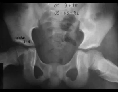
Eosinophilic granuloma can present with a clinical picture of fracture when it affects the spinal column where a wedging fracture of the vertebral body occurs, described as Calvé’s vertebra plano (fig. 38). In this situation, this injury may progress to spontaneous healing, and restoration of the vertebra body may even occur.

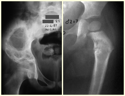
Other locations where micro-fractures can occur are when they affect the supra-acetabular region (fig. 39), or in load-bearing areas such as the proximal metaphyseal portion of the femur (fig. 40), due to cortical erosion. medial.  

37 – reação periosteal lamelar grossa em granuloma eosinófilo. Não há lesão extra-óssea. Fig. 38 – vértebra plana de Calvé (fratura acunhamento do corpo vertebral em granuloma eosinófilo).
37 – thick lamellar periosteal reaction in eosinophilic granuloma. There is no extra-osseous injury. Fig. 38 – Calvé plano vertebra (wedge fracture of the vertebral body in eosinophilic granuloma).
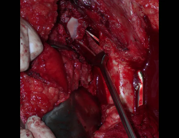
Figura 39 – granuloma eosinófilo do ilíaco – fratura afundamento do acetábulo. Fig. 40 – Fratura incompleta da cortical medial do fêmur, devido à granuloma eosinófilo.
Figure 39 – eosinophilic granuloma of the iliac – sinking fracture of the acetabulum. Fig. 40 – Incomplete fracture of the medial cortex of the femur, due to eosinophilic granuloma.

This lesion responds well to simple curettage surgical treatment, with the need to add a bone graft being exceptional.

Click here to read more.

Fibrous Dysplasia –

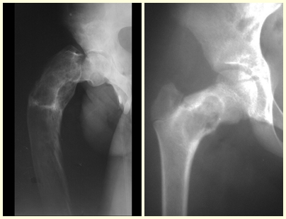
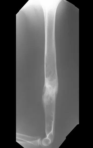
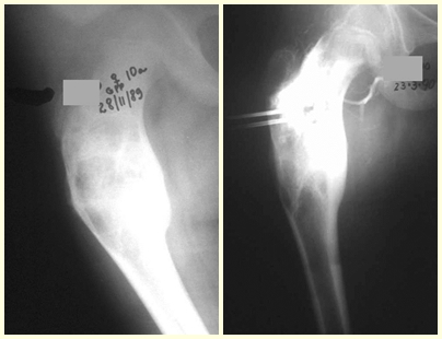
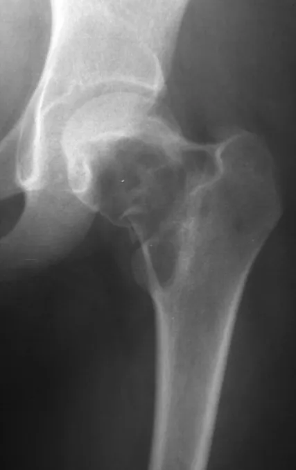
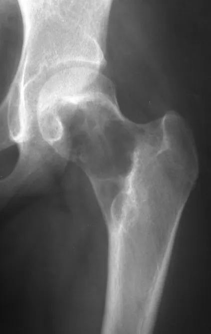
Fibrous dysplasia is a pseudo-tumor lesion that most frequently leads to bone deformity. However, when it affects the femur, it can cause a previous deformity, like a shepherd’s crook, characteristic of this condition, with consequent fracture (fig. 41). The femoral neck region with fibrous dysplasia often develops a fracture, even without previous deformity (fig. 42). 

To correct the defect, it is necessary to curettage the lesion, fill it with autologous bone graft and corrective osteotomies of the deformity (fig. 43). A fracture in this location can be difficult to resolve, due to the difficulty in consolidation due to the dysplastic appearance of the bone (fig. 44), leading to recurrence of the disease and deformity.

Figura 41 – Deformidade em cajado de pastor, seguida de fratura. Fig. 42 – fratura do colo femoral, em displasia fibrosa, sem deformidade prévia.
Figure 41 – Deformity in a shepherd’s crook, followed by fracture. Fig. 42 – fracture of the femoral neck, in fibrous dysplasia, without previous deformity.
Figura 43 – osteotomia corretiva, com enxerto autólogo. Fig. 44 – doença poli cística do colo femoral, por displasia fibrosa.
Figure 43 – corrective osteotomy, with autologous graft. Fig. 44 – polycystic disease of the femoral neck, due to fibrous dysplasia.
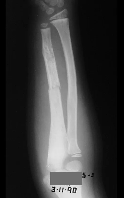
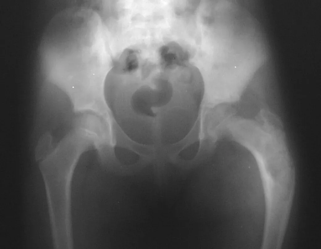
Fibrous dysplasia can be polyostotic (fig. 45) and be part of the MacCune-Albhright syndrome, characterized by fibrous dysplasia, precocious puberty and skin patches (fig. 46). Fractures can occur even without significant trauma, due to bone fragility (fig. 47).
Figura 45 – lesão no fêmur, por displasia fibrosa.
Figure 45 – injury to the femur, due to fibrous dysplasia.
Figura 46 – Mancha café com leite, em síndrome de MacCune-Albhright.
Figure 46 – Café-au-lait spot, in MacCune-Albhright syndrome.
Figura 47 – Fratura da ulna, devido à displasia fibrosa.
Figure 47 – Fracture of the ulna, due to fibrous dysplasia.
This disease tends to stabilize after puberty (fig. 48 and 49), but sometimes several surgical procedures are necessary during growth (fig. 50 and 51) to avoid compensatory deformities and achieve successful treatment. treatment.
48 – recidiva da deformidade, em síndrome da Albright .
48 – recurrence of the deformity, in Albright syndrome.
Figura 49 – criança em desenvolvimento com recidiva .
Figure 49 – developing child with relapse.
Figura 50 – nova recidiva da deformidade, necessidade de reoperação .
Figure 50 – new recurrence of the deformity, need for reoperation.
Figura 51 – estabilização da doença displásica após a adolescência .
Figure 51 – stabilization of dysplastic disease after adolescence.

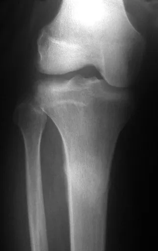
Fibrous dysplasia may also be part of congenital pseudoarthrosis, which most frequently affects the distal third of the tibia, but can occur in other locations such as the proximal third of the tibia (figures 52, 53 and 54), with all the difficulties in reaching it. if consolidation.

Figura 52 – Deformidade proximal da tíbia em displasia fibrosa. Fig. 53 – Fratura
Figure 52 – Proximal tibial deformity in fibrous dysplasia. Fig. 53 – Fracture
Figura 54 – Aspecto cirúrgico do foco displásico de pseudo-artrose, em displasia fibrosa.
Figure 54 – Surgical appearance of the dysplastic focus of pseudoarthrosis, in fibrous dysplasia.

Congenital pseudoarthrosis is a condition that deserves to be studied in a separate chapter.

Aneurysmal Bone Cyst –

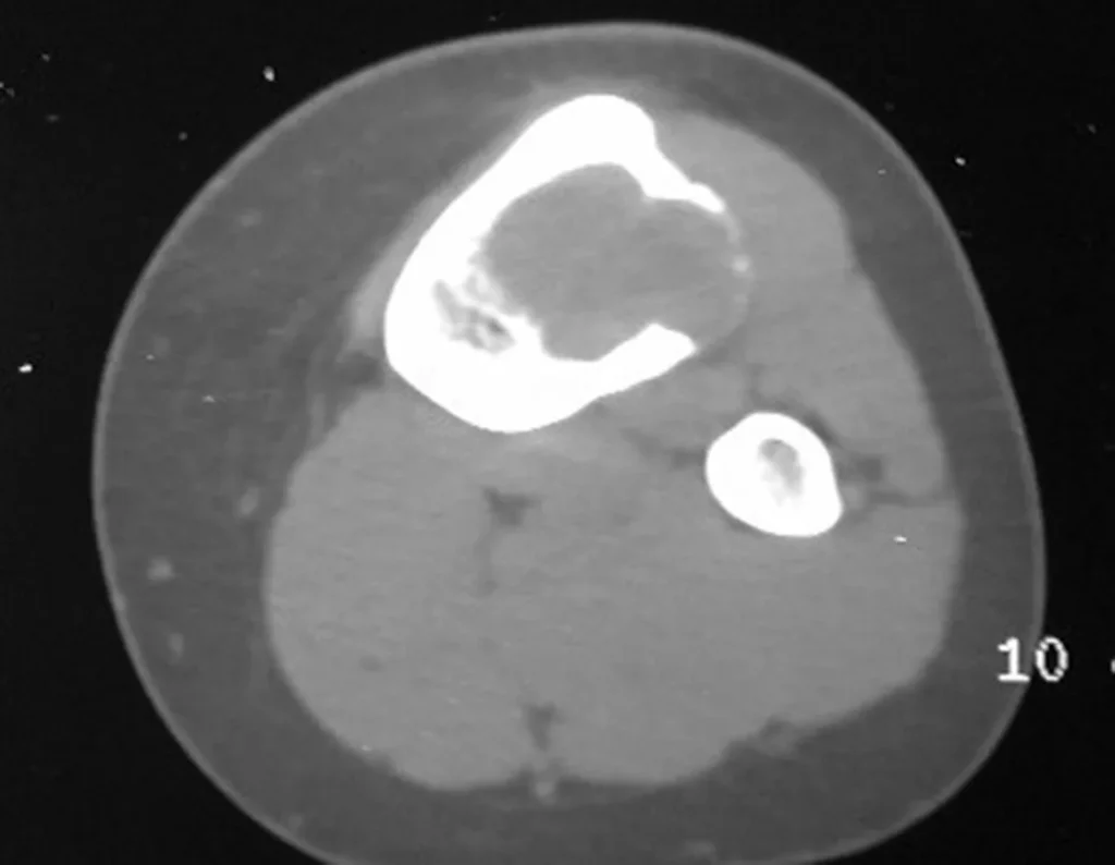
The aneurysmal bone cyst, also called multilocular hematic cyst, is a lesion of insufflative bone rarefaction filled with serosanguineous fluid, interspersed with spaces varying in size and separated by septa of connective tissue containing trabeculae of bone or osteoid tissue and ostoclastic giant cells ( figs. 55 and 56).

Figura Figura 55 – Cisto ósseo aneurismático da tíbia. As lojas ocorrem em número e tamanho variados, aglomerando-se e provocando erosão do trabeculado ósseo, que se expandem e insuflam a cortical.
Figure Figure 55 – Aneurysmal bone cyst of the tibia. The stores occur in varying numbers and sizes, clumping together and causing erosion of the bone trabeculae, which expand and inflate the cortex.
Figura Figura 56 – A tomografia revela área radiolucente; erosão óssea; afilamento da cortical e insuflação. sem focos de calcificação.
Figure Figure 56 – Tomography reveals a radiolucent area; bone erosion; cortical thinning and inflation. no foci of calcification.

The patient generally presents with mild pain at the site of the injury, when the affected bone is superficial, and inflammatory signs such as increased volume and heat may be observed. Generally, the patient correlates the onset of symptoms with some trauma.  

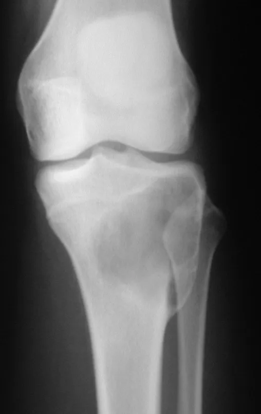
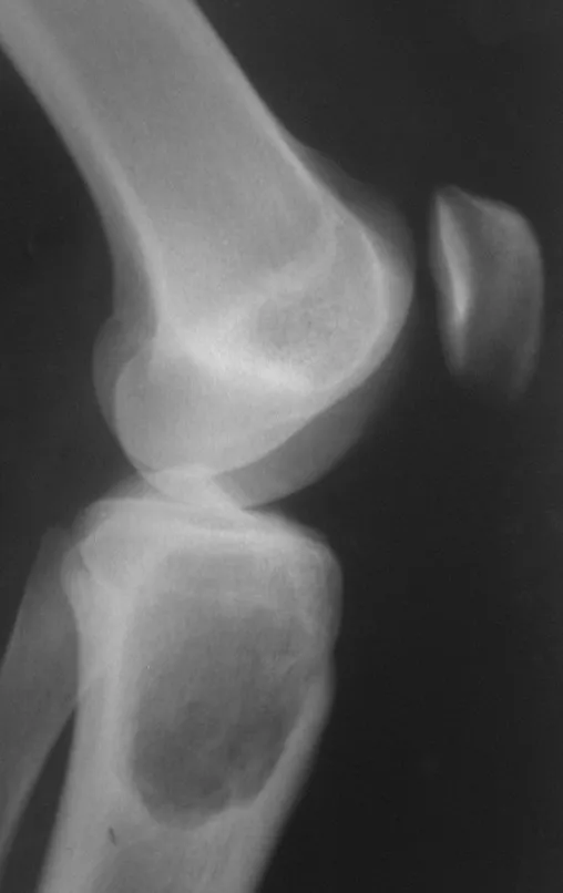
In evolution there may be a slow, progressive or rapidly expansive increase. It affects any bone, most frequently the lower limbs, tibia (figs. 57 and 58) and femur representing 35% of cases.

Figura Figura 57 – COA metafisário da tíbia com insuflação da cortical, erodindo a placa de crescimento.
Figure 57 – Metaphyseal AOC of the tibia with cortical inflation, eroding the growth plate.
Figura Figura 58 – aspecto homogênio com erosão da cortical
Figure 58 – homogeneous appearance with cortical erosion

The vertebrae are also affected by this injury, including the sacrum. In the pelvis, the iliopubic branch is most frequently affected. They can mimic joint symptoms when they reach the epiphysis. Compromise in the spine can cause compressive neurological symptoms, although in most cases it affects the posterior structures.

         The treatment of choice has been marginal resection or intra-lesional curettage, followed by filling the cavity with an autologous or homologous graft, when necessary. The cavity can also be filled with methylmethacrylate, although our preference is to use an autologous graft when possible, as it is a benign lesion. Some authors associate intralesional adjuvant treatment with the application of phenol, electrothermia or cryotherapy. In classic aneurysmal bone cysts, I do not see the point of this therapy, which, however, should be applied when the surgeon finds a “suspicious” area that was not detected on imaging. If the aforementioned benign tumors are involved, which may be accompanied by areas of aneurysmal bone cyst, local adjuvant therapy will be beneficial.

Some bone segments such as the ends of the fibula, clavicle, rib, distal third of the ulna, proximal radius, etc. can be resected, without the need for reconstruction.

In other situations, we may need segmental reconstructions with free or even vascularized bone grafts or joint reconstructions with prostheses in advanced cases with major joint involvement. In the spine, after resection of the lesion, arthrodesis may be necessary to avoid instability.

Radiotherapy should be avoided due to the risk of malignancy, however it may be indicated for the evolutionary control of lesions in difficult to access locations, such as the cervical spine, for example, or other situations in which surgical re-intervention is not recommended.

Embolization as an isolated therapy is controversial. However, it can be used preoperatively to minimize bleeding during surgery. This practice is most used in cases of difficult access, although its effectiveness is not always achieved. Infiltration with calcitonin has been reported with satisfactory results in isolated cases.

Recurrence may occur, as the phenomenon that caused the cyst is unknown and we cannot guarantee that surgery repaired it. The recurrence rate can reach thirty percent of cases.

Simple Bone Cyst –

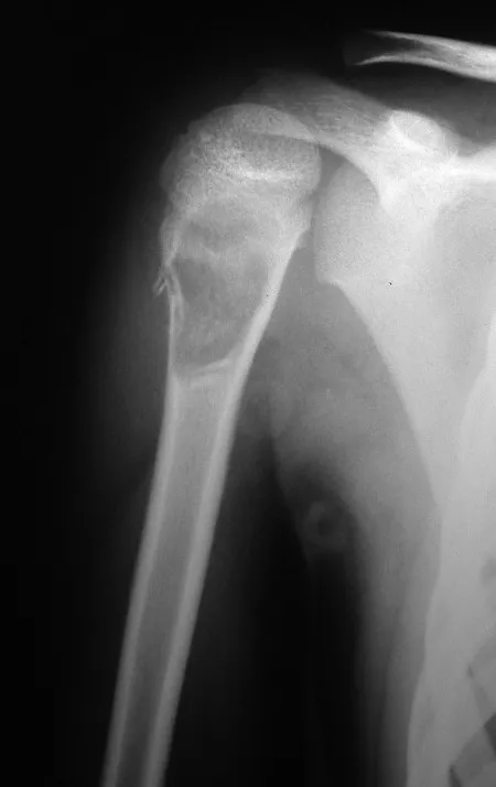
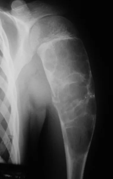
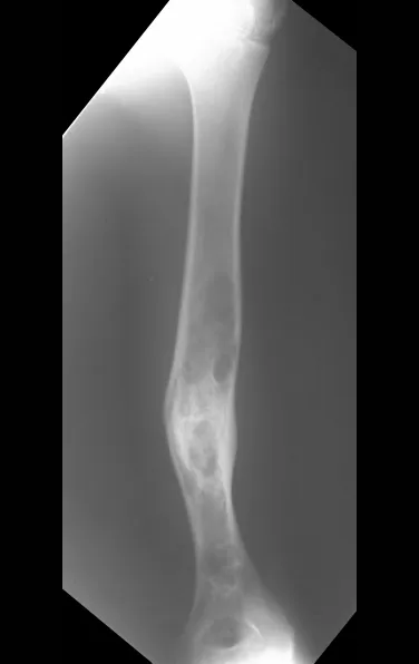
A simple bone cyst is a pseudo-tumor lesion that can occur in any part of the skeleton and most frequently presents with fracture (figures 59 to 64).

Figura 59 – fratura metafisária do úmero em C.O.S..
Figure 59 – metaphyseal fracture of the humerus in COS.
Figura 60 – microfratura do rádio em C.O.S..
Figure 60 – microfracture of the radius in COS.
Figura 61 – fratura infracção do acetábulo em C.O.S..
Figure 61 – Infraction fracture of the acetabulum in COS.
Figura 62 – Fratura completa do colo femoral em C.O.S..
Figure 62 – Complete fracture of the femoral neck in COS.
Figura 63 – Fratura do fêmur em C.O.S..
Figure 63 – Femur fracture in COS.
Figura 64 – Fratura da tíbia, após entorse em C.O.S..
Figure 64 – Tibial fracture, after COS sprain.

A simple bone cyst can occasionally be diagnosed due to an increase in volume, but when it presents a painful symptom, it is generally related to micro fractures or often a complete fracture.

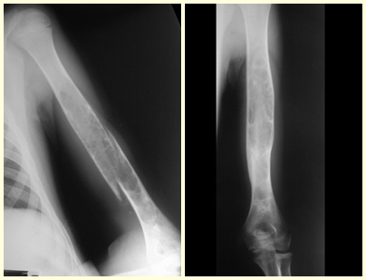
            The humerus is the most affected bone. Micro-fractures can eventually provide partial “cure” in some areas of the cyst and with growth the metaphysis moves away from the lesion, which begins to occupy the diaphyseal zone (fig. 65 and 66). This progression to the diaphysis can occur asymptomatically and a new painful clinical manifestation may occur acutely (fig. 67).

Figura 65 – Cisto ósseo metafisário do úmero.
Figure 65 – Metaphyseal bone cyst of the humerus.
Figura 66 – cicatrização da região metafisária, crescimento ósseo e deslocamento diafisário da lesão remanescente.
Figure 66 – healing of the metaphyseal region, bone growth and diaphyseal displacement of the remaining lesion.
Figura 67 – aumento da lesão e fratura.
Figure 67 – increase in injury and fracture.
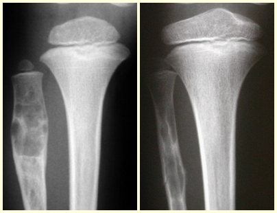
Bone cysts in older children and distant from the growth plate are considered mature cysts, which can heal with different treatment methods, including after the occurrence of a fracture (fig. 68 to 71).
Figura 68 – COS da fíbula. Fig. 69 – Fratura em COS.
Figure 68 – COS of the fibula. Fig. 69 – Fracture in COS.
Figura 70 crescimento ósseo metafisário e “migração” da lesão em direção à diáfise. Fig. 71 Cicatriz de COS que foi distanciando-se da placa de crescimento que teve cura expontânea.
Figure 70 metaphyseal bone growth and “migration” of the lesion towards the diaphysis. Fig. 71 COS scar that distanced itself from the growth plate that healed spontaneously.

In these situations, the treatment adopted must be appropriate for the bone and the fracture in question, and may be closed or open, with the indication of filling with a bone graft depending only on the specific needs of the fracture, when surgical treatment is indicated. 

In mature bone cysts, the complete fracture causes great decompression of the lesion and consolidation and healing of the lesion can be achieved simultaneously. However, in some cases, there is a need for additional treatment of the cyst, after consolidation of the fracture, when closed treatment is chosen (fig. 72 to 78).

Figura 72 – Fratura diafisária do úmero, em cisto ósseo maduro (cisto distante da placa de crescimento em adolescente). Fig. 73 – Consolidação após imobilização com tipóia.
Figure 72 – Diaphyseal fracture of the humerus, in a mature bone cyst (cyst distant from the growth plate in an adolescent). Fig. 73 – Consolidation after immobilization with a sling.
Figura 74 – Refratura. Fig. 75 – Novo tratamento incruento.
Figure 74 – Refracture. Fig. 75 – New bloodless treatment.
Figura 76 – Consolidação e persistência de áreas císticas.
Figure 76 – Consolidation and persistence of cystic areas.
Figura 77 Calo de fratura e áreas císticas.
Figure 77 Fracture callus and cystic areas.
Figura 78 – Função após múltiplas fraturas.
Figure 78 – Function after multiple fractures.
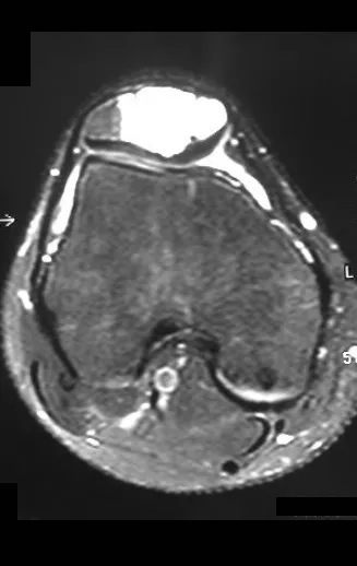
In our infiltration technique, we usually evaluate the cavity by injecting contrast, aiming to verify whether the cyst is unicameral or whether it has septa forming gaps that would require an individual approach. We began to observe through radioscopy the presence of contrasted vascular flow when contrast was injected into the cyst. We believe that there is an intraosseous pseudo-aneurysm that, when swirling, causes cystic erosion (fig. 79 to 86).
Figura 79 – Diagnóstico do cisto após fratura proximal do fêmur. Fig. 80 – Tratamento incruento, com tração cutânea por seis semanas
Figure 79 – Diagnosis of the cyst after a proximal femur fracture. Fig. 80 – Bloodless treatment, with skin traction for six weeks
Figura 81 – Consolidação da fratura, com cura parcial da lesão cística. Fig. 82 – Tratamento do cisto remanescente com infiltração. Observa-se contraste evidenciando a presença vascular na fisiopatologia desta lesão.
Figure 81 – Consolidation of the fracture, with partial healing of the cystic lesion. Fig. 82 – Treatment of the remaining cyst with infiltration. Contrast is observed, highlighting the vascular presence in the pathophysiology of this lesion.
Figura 83 – Cisto ósseo consolidado, após fratura e tratamento com infiltração. Presença de lesões císticas remanescentes.
Figure 83 – Consolidated bone cyst, after fracture and infiltration treatment. Presence of remaining cystic lesions.
Figura 84 – COS da patela, corte sagital.
Figure 84 – COS of the patella, sagittal section.
Figura 85– cisto unicameral da patela, corte axial.
Figure 85– unicameral cyst of the patella, axial section.
Figura 86 – contraste demonstra a alteração vascular, com verdadeiras fístulas arterio- venosas na fisiopatologia.
Figure 86 – contrast demonstrates the vascular alteration, with true arteriovenous fistulas in the pathophysiology.
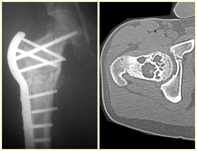
Some locations, such as the femoral neck, deserve special attention and should preferably be operated on before a fracture occurs (fig. 87 and 88).
Figura 87 – Cisto ósseo simples no colo femoral, antes da fratura. Fase de tratamento mais simples
Figure 87 – Simple bone cyst in the femoral neck, before fracture. Simplest treatment phase
Figura 88 – Fratura do colo femoral em cisto ósseo
Figure 88 – Fracture of the femoral neck in a bone cyst
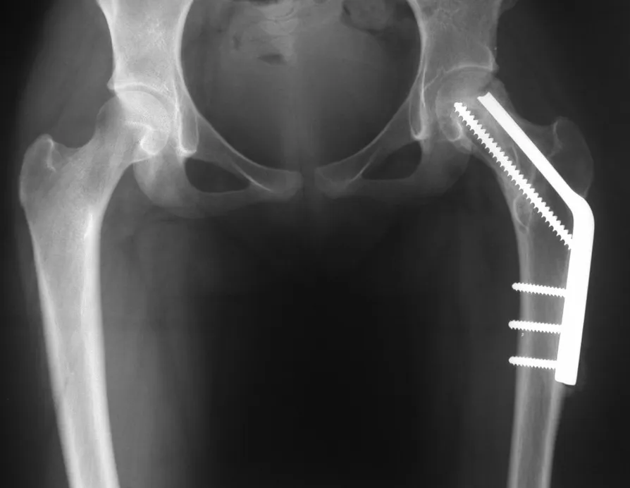
Treating this injury after a fracture requires specific and complex planning to be successful (fig. 89 to 94).
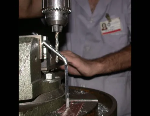
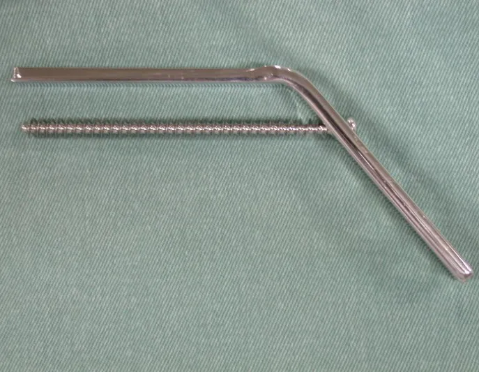
Figura 89 – Preparo do orifício da placa angulada.
Figure 89 – Preparation of the angled plate hole.
Figura 90 – Orifício adaptado para permitir orientação do parafuso paralelo à lâmina.
Figure 90 – Hole adapted to allow screw orientation parallel to the blade.
Figura 91 – Preparação do enxerto.
Figure 91 – Graft preparation.
Figura 92 – redução da fratura, posicionamento do enxerto e fixação.
Figure 92 – fracture reduction, graft positioning and fixation.
Figura 93 – pós-op um ano.
Figure 93 – one year post-op.
Figura 94 – rx perfil. Fig. 95 – função após 1 ano.
Figure 94 – profile x-ray. Fig. 95 – function after 1 year.

Click here to see the full case

Stress Fracture –

 Stress fractures deserve special attention in this article both because they are more frequent than reported in the literature, as many cases go unnoticed, and because of the florid appearance that imaging studies portray, causing difficulty in differential diagnosis.

The child complains of pain, usually after physical exertion, which, as it is mild, ends up resolving spontaneously.

However, an orthopedist may be consulted and, when requesting an x-ray, be surprised by a periosteal reaction in the metaphyseal region in a growing patient.

The concern about the possibility of osteomyelitis, eosinophilic granuloma, osteosarcoma or Ewing’s sarcoma is justified, but it is necessary to be aware of clinical aspects, such as time of evolution, improvement factors, local appearance, so as not to complicate this diagnosis, which is  clinical. radiological  (fig. 96 and 97).

Figura 96 – dor há um mês .
Figure 96 – pain for a month.
Figura 97 – reação periosteal.
Figure 97 – periosteal reaction.
Carrying out other tests such as bone mapping (fig. 98) and computed tomography (fig. 99) confirm the existence of the lesion, but may not be sufficient to clarify the diagnosis.
Figura 98 –hipercaptação.
Figure 98 – hypercapture.
Figura 99 – TC com produção de osso
Figure 99 – CT with bone production

It is necessary to evaluate and ask:  in the time it took to carry out these tests, was there no clinical improvement?

Magnetic resonance imaging is an exam that needs to be interpreted very carefully, as the fracture causes intra- and extra-osseous edema that can scare less experienced people.

We must observe the detail of the two low signal points of the fracture callus in the lateral and medial cortex in figure 100 as well as the low signal point of the bone callus in the posterior cortex in figure 101.

Figura 100 – RM em fratura de stress.
Figure 100 – MRI in stress fracture.
Figura 101 – Baixo sinal do calo ósseo
Figure 101 – Low signal from bone callus

The inflammatory process of the fracture, with marked hemorrhage and edema, is extensive. The histology of the fracture callus may mimic osteosarcoma. There is a known case of amputation due to an erroneous diagnosis of osteosarcoma in a patient with a stress fracture.

            Observation for two to three weeks is essential for an accurate diagnosis and is not considered bad practice, even in neoplasms. The x-ray taken three weeks later showed the stress fracture (fig. 102 and 103) and the clinical picture with improvement in symptoms and reduction in edema reaffirms the diagnosis. The clinic is sovereign.

Figura 102 – Rx após 3 semanas.
Figure 102 – Rx after 3 weeks.
Figura 103 – A linha da fratura é nítida.
Figure 103 – The fracture line is clear.

Author: Prof. Dr. Pedro Péricles Ribeiro Baptista

 Orthopedic Oncosurgery at the Dr. Arnaldo Vieira de Carvalho Cancer Institute

Leave a Reply

Hello! How can we assist you?